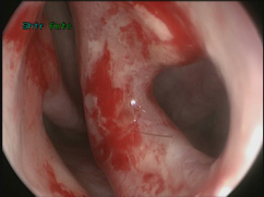
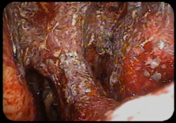
Read more about the article Saddle Nose Deformity and Septal Perforation:  Correcting Nasal Collapse
Figure 4a,b. Nasal reconstruction of anterior septal perforation. Patient also complained that her nose was droopy and wide after developing the perforation. Pre and post-operative photos demonstrate improvement in tip projection after reconstruction of the septum and rhinoplasty.

Saddle Nose Deformity and Septal Perforation: Correcting Nasal Collapse

Nasal Septum Repair/Septal Perforation Surgery/ Saddle Nose Deformity Jason S. Hamilton, MD, FACS Director of the Division of Plastic and Reconstructive Surgery Osborne Head and Neck Institute, Los Angeles, CA…

Continue ReadingSaddle Nose Deformity and Septal Perforation: Correcting Nasal Collapse
Read more about the article Complications of Ear Piercing: Perichondritis and Ear Disfigurement
Figure 1: Perichondritis of the right ear that has progressed to chondritis (right). Note the discoloration, inflammation, and grainy appearance of the skin compared to the normal left ear (left).

Complications of Ear Piercing: Perichondritis and Ear Disfigurement

  Body piercing has become more popular and less taboo in recent years. With an increase in piercing there has been a concomitant increase in complications .One area particularly at…

Continue ReadingComplications of Ear Piercing: Perichondritis and Ear Disfigurement
Read more about the article Options After Unsuccessful Sialendoscopy
Figure 1. Salivary gland stones counted and measured for display after removal from the ductal system via a sialendoscopy procedure performed at the Osborne Head and Neck Institute. After salivary stones are removed they are correlated with stones visualized on a preoperative CT scan.

Options After Unsuccessful Sialendoscopy

Can I avoid surgery after a failed sialendoscopy? Question: I have a long history of salivary gland stones and conventional treatments have become ineffective. Recently I developed a large stone…

Continue ReadingOptions After Unsuccessful Sialendoscopy
Read more about the article Orbital Floor Blowout Fracture – Treatment Options
Figure 2a,2b. An example of a patient presenting with a right orbital floor blowout fracture. Bruising and limited eye movements secondary to swelling are common clinical presentations (top). CT scan demonstrates common findings of a blow out fracture with evidence of a depressed right orbital floor (bottom).

Orbital Floor Blowout Fracture – Treatment Options

  What are my treatment options for an orbital blowout fracture? Question: I currently play baseball for my local university and was injured during our last game a few days…

Continue ReadingOrbital Floor Blowout Fracture – Treatment Options