
- Hearing Restoration Surgeries for Congenital Microtia - September 4, 2019

- Risks of Surgery for Surfer’s Ear - October 26, 2017
- The Dangers of Untreated Exostosis / Surfer’s Ear - October 26, 2017
- Surfer’s Ear Treatment: Chisel vs Drill - October 26, 2017
Overview
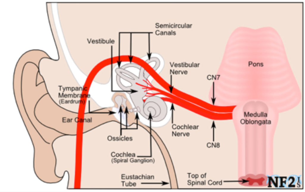
Congenital microtia is a deformity of the outer ear, but it can also impact structures inside the ear. Patients with congenital microtia may present with canal stenosis (narrowing of the ear canal) or aural atresia (absence of the ear canal). In these cases, a canalplasty may be performed to widen or create an ear canal. In addition, patients can lack a tympanic membrane (eardrum), or only have a partial one, which can be treated with a surgical procedure called a tympanoplasty. Microtia patients can also have problems with their ossicles (malleus, incus, and stapes) which may be too small, deformed, or absent. In these cases, an ossicular chain reconstruction would be recommended. These problems can occur independently of another, but it is common for patients to have more than one issue. Whether patients present with only one issue or all three, they will have hearing loss because the ear canal, tympanic membrane, and ossicles each play a role in hearing; surgical treatment would help restore hearing.
Canalplasty
Canalplasty is a surgical procedure to treat partial or complete obstruction of the external auditory canal (ear canal) from bone or skin. In patients with canal stenosis (narrowed canal), the procedure widens the ear canal, which allows more sound to reach the tympanic membrane (eardrum). In patients with aural atresia (complete canal closure), canalplasty creates an ear canal. The surgery involves drilling or chiseling through the bone that is obstructing the canal until the tympanic membrane is reached. Canalplasty is a highly effective procedure, but it does carry significant risks. In rare cases, complete hearing loss, infection, and harm to other ear structures may occur. One of the most significant concerns when performing a canalplasty is the integrity of the facial nerve. The facial nerve runs through the external auditory canal and is resposible for motion of the face. This nerve is at risk for damage in canalplasty, resulting in facial nerve paralysis. The surgeon must carefully and often use nerve identification techniquest to protect the nerve. However, because the patient has abnormal anatomy, the facial nerve is often in an atypical location and may be therefore injured. A CT scan prior to surgery can help with surgical planning so that the location of the nerve, as well as the anatomy of the canal and other ear structures, can be assessed. In cases where facial nerve anatomy is not favorable, canalplasty not be recommended; a bone-anchored hearing aid (BAHA) may be recommended for the patient.
Tympanoplasty
Tympanoplasty is surgical procedure that is performed when microtia patients have a deficient or absent eardrum. Fascia, a tissue found between skin and muscle, is taken from behind the patient’s ear. It is used as a graft to replace or create the missing portion of the eardrum. If a sufficient ear canal exists and the graft needed is fairly small, the surgeon may choose a transcanal approach, which involves placing the graft through the ear canal, without external incisions. In more complicated cases, the surgeon may make an incision behind the ear, which allows access for more involved surgery. This is called a post-auricular approach. As with canalplasty, there is risk of facial nerve injury in a tympanoplasty, but occurs more infrequently. There are also chances of permanent hearing loss and altered sense of taste, but these are rare.
Ossicular Chain Reconstruction
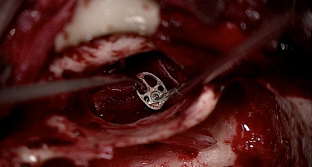
Ossicular chain reconstruction is a surgical procedure to replace the ossicles with a prosthesis, and is used in patients where ossicles are underdeveloped or absent. The ossicles are normally found in the middle ear, behind the eardrum, so the eardrum must be lifted to access them. Risks of this procedure include facial nerve injury, hearing loss, or a dislodged prosthesis, although these are all uncommon.
