Salivary Gland Stone removal
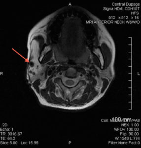
Figure 1: Axial MRI of the head demonstrating an arteriovenous malformation (arrow).
The generally inaccessible locations of the salivary glands have traditionally led to challenges in accurate diagnosis and treatment of gland pathology. Although greatly improved in modern times, clinicians continue…
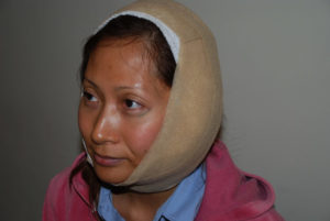
Figure 1: Special compressive dressings are placed temporarily during healing to reduce the likelihood of fluid leakage into surrounding tissues and subsequent sialocele formation.
Can sialocele be prevented when undergoing salivary gland surgery? Question: I have been evaluated for a salivary gland mass by a head and neck surgeon and he has recommended…
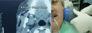
Figure: Left - CT scan of the head with a visible stone in the right parotid salivary gland. Right – Lithotripsy patient undergoing procedure.
Is lithotripsy an option for the treatment of salivary stones? Question: My ENT doctor diagnosed me with “sialolithiasis”, a salivary gland stone. Before my diagnosis, I periodically noticed that…
How do I know if I’m a candidate for Sialendoscopy of my salivary gland stones? Question: I just got back from the ENT. I had a CAT scan of…
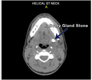
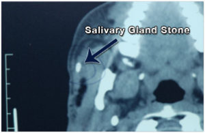
Figure 1. Salivary Gland Stone in left submandibular gland.
How are salivary gland stones diagnosed? Question: I had a consultation for swelling in my right cheek that comes and goes when I eat sour foods. So times it is…
Dr. Ryan Osborne, Director of Head & Neck Surgery and Salivary Gland Disorders, at the Osborne Head & Neck Institute, specializes in the non-surgical removal of salivary gland stones causing…
American Health Front (AHF) recently interviewed Dr. Ryan F. Osborne, Director of the Osborne Head and Neck Institute, about advances in parotid tumor treatment. Although relatively a small number of…

Dr. Ryan Osborne,
View Dr. Ryan Osborne's television appearance on The Doctors below: Recently, Dr. Ryan F. Osborne, Director of the Division of Head and Neck at the Osborne Head and Neck Institute,…


