- Question: How do the ingredients in e-cigarettes and vaporizers affect respiratory health? - August 16, 2019
- Bad Technique and Vocal Injury - January 9, 2019
- Is Edible Marijuana Dangerous for the Voice? Myths Dispelled - December 18, 2018
- Surprise! You have a hemorrhage - January 31, 2018
- Graves’ Disease: Treatment Overview - September 25, 2017
- Adele and the Stigma of Vocal Injury - July 11, 2017
- Vocal Curbside Consult: How does the thyroid affect the voice? - May 16, 2017
- Vocal Curbside Consult: How do hormones affect the voice? - May 3, 2017
- Vocal Curbside Consult: How do emotion and stress affect the voice? - April 17, 2017
- Vocal Curbside Consult: Vocal Recovery After Illness - April 7, 2017
The patient is a 26 year old professional singer who has noted significant postnasal drip since performing in a smoky venue 3 weeks earlier. She feels that something is “stuck” in her throat and that singing is more fatiguing than usual. She also notes an increasingly frequent need to clear her throat. This temporarily improves her voice, however, the troubling sensation eventually returns. She denies loss of vocal range or change in vocal quality, noting that she has always had a raspy voice. She has no significant medical history and no history of seasonal or perennial allergies.
Her general physical exam was non-contributory and she did not demonstrate any signs of postnasal drip.
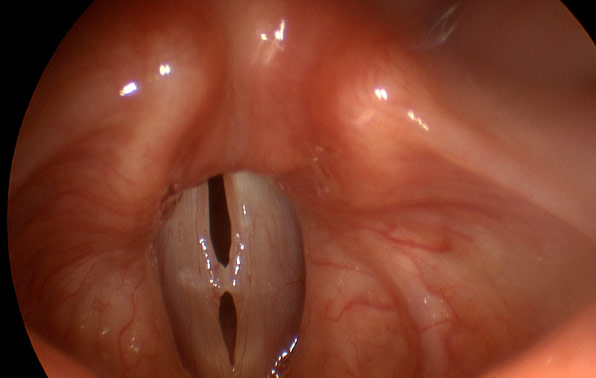
Videostroboscopy was performed:
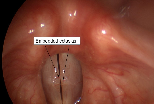
Stroboscopy demonstrates bilateral vocal fold polyps within the striking zone. There are large embedded blood vessels in the polyps, suggesting they formed due to vocal hemorrhage.
This patient has bilateral vocal fold polyps. However, she denies any change in vocal range or vocal quality. She only notes “throat clearing” and has a triggering event. This brings to light the critical nature of a screening vocal exam for establishing a baseline or reference level of vocal health. Had this patient been screened when feeling normal, and the presence of the polyps noted, the polyps would not be suspected in contributing to her current symptoms. However, without the screening exam, her history of singing in an environmentally unfavorable setting and subsequent polyp formation, cannot be definitively ruled out and must be considered as possible causes of her symptoms.
Context is critical in cases of vocal injury. Treatment of this patient hinges almost entirely on whether we believe these polyps to be new and causative of symptoms or old and unrelated to her complaints. Because vocal polyps are a reasonably serious vocal injury, they will be considered and treated first in the short term as if they were acute. Based on her response to conservative treatment, further treatment will be customized and initiated. A prior screening exam, performed when the patient felt vocally healthy, would have potentially eliminated any diagnostic uncertainties and made the course of treatment completely clear.
To learn more about Dr. Reena Gupta or vocal screening exams, please visit www.voicedoctorla.com.


