
- Risks of Surgery for Surfer’s Ear - October 26, 2017
- The Dangers of Untreated Exostosis / Surfer’s Ear - October 26, 2017
- Surfer’s Ear Treatment: Chisel vs Drill - October 26, 2017
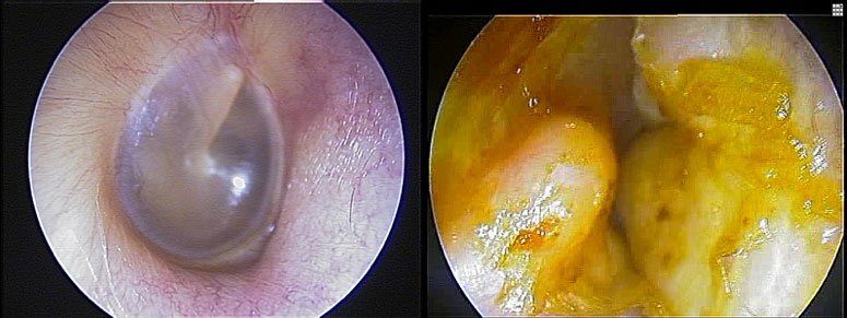
Surfer’s ear, also known as exostosis, is an abnormal growth of bone inside the ear canal. These growths produce symptoms such as trapping of water and wax, which can lead to infections and hearing loss. Surfer’s ear can become quite bothersome for the daily life of a surfer or swimmer, as water, sand, and debris can get stuck in the ear after almost every water activity.
Many surfers will opt not to have surgery due to the risks. Further, they become accustomed to symptoms over several years, as the exostoses grow slowly. They may not perceive the loss of hearing or other associated symptoms because symptoms occur gradually. However, as exostoses become larger from continued cold water exposure, the surgery becomes increasingly complicated. While it is never advisable to rush into surgery, it is important to become educated about the real risks.
Exostosis surgery has a quicker recovery and higher success rate when done early, before exostoses become very large. Consulting with an otologist (and ENT with specialty training in ear disorders) is advised to ensure you are not ignoring a problem that can be easily treated and to prevent complications of more advanced disease.
What are the risks of surfer’s ear surgery?
In order to understand the risks of surgery, it is important to know the anatomy of the ear canal. The ear canal is surrounded by many important structures:
- Tympanic membrane (ear drum)
- Temporomandibular joint (jaw joint)
- Facial nerve
- Chorda tympani (nerve for taste)
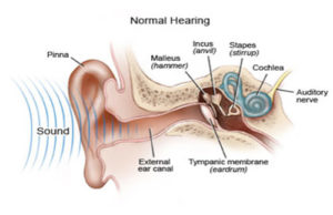
- Ossicles (bones that conduct hearing)
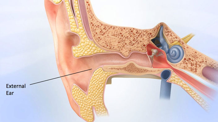
The ear canal is a very narrow structure, requiring the use of a microscope to perform surgery without damaging these vital structures.
Complications from exostosis and other canal surgery usually arise from surgeon inexperience or from extremely large or numerous exostoses. These may include:
- Poor skin healing inside the canal
- Damage to the ear drum
- Failure to restore normal ear canal contour and function
- Difficulty chewing
- Pain with chewing
The surgeon must use caution to protect the bone around the temporomandibular joint, as excessive removal can cause exposure of the joint. This can cause pain and difficulty with
chewing which is very difficult to treat. It is only through extensive experience with this surgery and specialty training that such complications can be avoided.
How likely are these complications?
These complications are very rare in the hands of a trained otologist (ear surgeon). There is higher risk of injuring these structures when exostoses are very severe. Large exostoses make landmarks difficult to identify and limit the space in which the otologist can operate. It is therefore important to ensure that your surgeon has significant experience working on patients with severe exostoses. Many of these complications are difficult to treat, making it critical to avoid these problems. Seek consultation with an experienced otologist to ensure the highest success rate with exostosis surgery.
To learn more about exostoses or surfer’s ear, please visit: www.eardoctorla.com
