- The Rare Case of Microtia - September 26, 2016
- Folded Ear in Newborns: Treatment Options - April 11, 2018
- Newborn Ear Deformity: What Can Be Done? - April 11, 2018
- Ear Molding: An Overview - November 2, 2017
- Otoplasty for Protruding Ears - September 26, 2017
- Basal Cell Carcinoma: Facial Reconstruction Timing - September 26, 2017
- Clinical Considerations of Mohs Reconstruction of Cheek Defect - September 26, 2017
- Basal Cell Carcinoma: Nasal Bridge Reconstruction - September 26, 2017
- Skin Cancers Involving the Eyebrow: Clinical Considerations - October 3, 2016
- Treatment of Multiple Skin Cancer Lesions - June 1, 2016
- Skin Cancer: Nasal Reconstruction and Scar Management - June 1, 2016
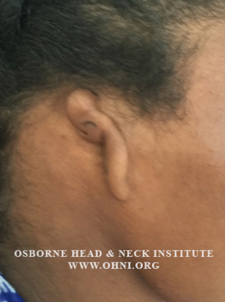 What is Microtia?
Microtia is a congenital condition that results in the deformation of the external ear. Microtia is relatively rare and occurs in 3 out of every 10,000 births. Treatment options have traditionally been a challenge for surgeons around the world. The difficulty in treating this condition resides in the complex structure of the ear and the task of creating an ideal anatomic environment for the new ear. Adding to the complexity, hearing can also be affected in microtia cases where the ear canal is involved. There are however several different grades of microtia that vary in severity. Microtia can be graded based on the Weerda Classification, which categorizes Microtia into four areas of increasing severity. The most severe form of microtia which is Grade 4 or Anotia is characterized by a complete absence of an ear. Grade 3 microtia is the most common form of microtia and is characterized by absence of the ear and the presence of a small malformation. There are several treatment options for microtia.
What treatments are available for microtia?
Autologous Reconstruction Using Rib Cartilage
This treatment involves carving and assembling a framework of cartilage harvested from the ribs and inserting the structure behind the deformed ear. The framework base is carved from the 6th and 7th rib cartilage using scalpels. The 8th rib is split to serve as the helix of the ear. The framework is created smaller than the unaffected ear to account for skin thickness. The skin pocket is made by a small incision anterior to the microtia vestige. Dissection over the mastoid allows skin to drape over the framework. The framework is then put into the pocket. 6 to 8 weeks after the initial surgery the earlobe is transposed into its proper position. Next, the framework is elevated from the mastoid, allowing the ear to appear more natural.
Reconstruction using Alloplastic Grafts
Another option is to use alloplastic grafts, implantable material such as Medpor or Stryker, to reconstruct the ear. These substances are stable, inert, and easily integrate with the patient’s tissue. Alloplastic grafts are also thermoplastic which allows them to mold to their surroundings. First, a scalpel is used to make an incision in the dermal papillae of the hair allowing for the removal of the deformed ear’s cartilage remnant. The reconstructed ear is then implanted into this same area, sutured into place, and covered with skin. The earlobe is transposed three months after the initial procedure.
What risks are involved with treatment of microtia?
Treatment of microtia can be associated with rare complications. During rib cartilage harvesting, the patient is at risk for a pneumothorax, a treatable condition that can result in a collapsed lung. Bruising, hematoma formation, and infection are also rare but possible complications. Use of Alloplastic Grafts during reconstruction is favored by some because they can be used on younger patients, decreases operation time, and eliminate the need for harvesting cartilage from the patient’s ribs. Alloplastic material also tends to be more aesthetically accurate. It should be noted that there is also a slight risk of infection with alloplastic techniques.
 What treatments are available for hearing loss?
Microtia can result in the absence of an ear canal and result in conductive hearing loss. Hearing in these cases can be improved by using the following methods:
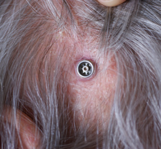
Hearing Aids
There are several options when it comes to selecting the right hearing aid. Bone Anchored Hearing Aids or BAHA, are attached to the skull by an implant that is placed by an otologist. These devices pick up sound waves and transmit sound through bone to the middle ear by small vibrations. Another option is to get an implantable hearing aid which attaches to the small bones of the middle ear. These aids work by moving the bones directly to produce sound.
Ear Canal Reconstruction
This is a surgical procedure to reconstruct and realign the ear canal to restore some of the natural hearing in a microtia patient. The goal of this surgery is to provide the patient with a clean, skin-lined canal and improve hearing.
All cases of microtia should be evaluated and treated by a qualified facial plastic surgeon and otologist. Otology and facial plastic surgery are subspecialties of otolaryngology. These specialists have specialized training and can address not only the cosmetic/aesthetic concerns of microtia but also the functional considerations.
To learn more about microtia and available treatments, please visit: www.eardoctorla.com.
